25 Sept 2026 · Case #020
Giant cell arteritis (temporal arteritis)
This page works the case through in full, answer included. If you would rather reason it out yourself: Play this case
Diagnosis
Giant cell arteritis (temporal arteritis)
First-line treatment
Immediate high-dose glucocorticoids
How the diagnosis is reached
New-onset headache over 50 with scalp tenderness and a markedly raised ESR is giant cell arteritis until disproven, and the jaw claudication settles it: pain that comes on with chewing and resolves with rest is the single most specific symptom, reflecting ischemia of the masseter through branches of the external carotid. The pulseless temporal artery on one side with a normal vessel opposite is the characteristic finding, and the girdle stiffness is the polymyalgia rheumatica that coexists in roughly half of cases; a normal creatine kinase distinguishes this from an inflammatory myopathy, and the raised alkaline phosphatase is the mild cholestatic picture of active disease, not liver or bone pathology. The episode of transient monocular visual loss is amaurosis fugax from involvement of the posterior ciliary arteries, and untreated it heralds permanent, painless and irreversible blindness, which can arrive within days and may then take the second eye. Ultrasound showing a non-compressible halo supports the diagnosis, and the thickened right axillary artery marks this as a large-vessel vasculitis reaching well beyond the temple. Biopsy is confirmatory, but the critical management principle is that high-dose glucocorticoids must be started the moment the diagnosis is suspected, without waiting for either. Because she has already had amaurosis fugax, that means intravenous methylprednisolone rather than the oral prednisolone which would serve for headache alone. Biopsy stays informative for about two weeks after steroids begin, and the skip lesions here explain why specimen length matters and why a negative biopsy does not exclude the disease. Tocilizumab is added as a steroid-sparing agent given the long treatment course and the burden of steroid toxicity in this age group, with bone and gastric protection alongside it for the same reason.
Key clinical clues
- New headache over age 50 with scalp tenderness in a new distribution
- Jaw claudication, pain on chewing that resolves with rest, the most specific symptom
- Thickened, tender, pulseless temporal artery with a normal contralateral vessel
- ESR 96 with thrombocytosis and normocytic anemia; normal CK despite girdle stiffness
- Transient monocular visual loss, do not wait for biopsy before starting steroids
The case in full
Presenting complaint
Patient history
A 73-year-old woman has three weeks of a new right-sided headache over her temple, different from her usual headaches. Brushing her hair on that side hurts. Her jaw aches after a few minutes of chewing and settles when she stops, so she avoids meat.
Examination and observations
Physical examination
The right temporal artery is thickened, tender and pulseless; the left pulsates normally. She has marked stiffness and aching in both shoulders and hips, worst in the hour after waking, but no true weakness. Fundoscopy and acuity are normal, with no scalp rash.
| Parameter | Value | Reference |
|---|---|---|
| Temperature | 37.6 °CRef 36.1–37.5 | 36.1–37.5 |
| Blood pressure | 142/78 mmHg | – |
| Heart rate | 84 bpmRef 60–100 | 60–100 |
Inflammatory markers and blood count
Laboratory results
A blood count and inflammatory markers are sent.
| Parameter | Value | Reference |
|---|---|---|
| ESR | 96 mm/hRef <30 | <30 |
| C-reactive protein | 84 mg/LRef <5 | <5 |
| Platelets | 612 ×10⁹/LRef 150–400 | 150–400 |
| Hemoglobin | 10.4 g/dLRef 12.0–15.5 | 12.0–15.5 |
| Mean cell volume | 87 fLRef 80–100 | 80–100 |
| Alkaline phosphatase | 184 U/LRef 44–147 | 44–147 |
| Creatine kinase | 82 U/LRef 30–200 | 30–200 |
Clinical course
Clinical course
While awaiting outpatient review two days later, she has a sudden episode in which the vision in her right eye greys out completely, as though a curtain had descended. It returns fully after about five minutes. She has had no weakness, speech disturbance or facial droop.
Vascular ultrasound
Imaging
Colour Doppler of both temporal arteries shows hypoechoic, concentric, non-compressible wall thickening around the lumen of the right superficial temporal artery over roughly 2 cm. The left shows a shorter similar segment, and the right axillary artery is also thickened.
Histopathology
Pathology
A 2 cm biopsy specimen is taken; a high-power H&E section through the vessel wall is shown below. The changes are patchy along the specimen, with several histologically normal segments between the affected areas.
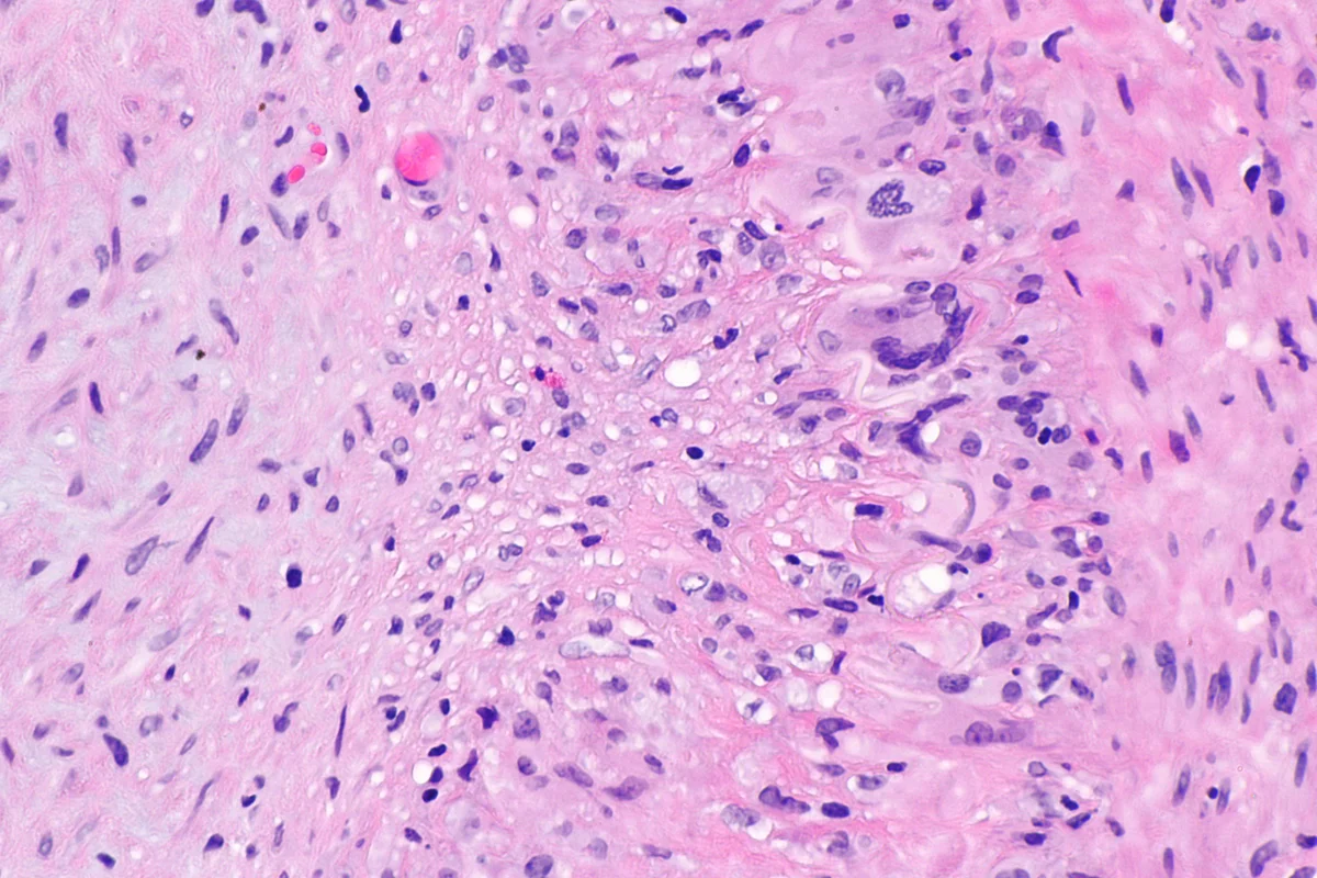
Temporal artery biopsy, H&E, high power.
Image by Nephron, via Wikimedia Commons. Licence: CC BY-SA 3.0.