24 Sept 2026 · Case #019
Multiple myeloma
This page works the case through in full, answer included. If you would rather reason it out yourself: Play this case
Diagnosis
Multiple myeloma
First-line treatment
Bortezomib-based induction (bortezomib, lenalidomide and dexamethasone)
How the diagnosis is reached
Back pain that wakes a 68-year-old at night and no longer answers to paracetamol is not mechanical, and the constipation, exhaustion and afternoon muddle beside it are one complaint rather than three: a calcium of 12.4 mg/dL. With the anemia and a creatinine of 2.3 mg/dL, the CRAB tetrad is complete. Several details then separate myeloma from its mimics. A normal alkaline phosphatase alongside extensive lytic disease is characteristic: myeloma lesions come from osteoclast activation with osteoblasts suppressed, so there is no reparative bone formation to raise the ALP or light up an isotope scan. That is why the negative scan misleads and why skeletal survey or whole-body CT is the right imaging; metastatic carcinoma provokes an osteoblastic reaction and is usually scan-positive. A suppressed parathyroid hormone excludes primary hyperparathyroidism as the cause of hypercalcemia. The very high total protein with low albumin, marked rouleaux and an ESR over 100 all reflect the circulating paraprotein. Recurrent infections follow from immunoparesis, visible here as suppressed IgA and IgM. The diagnosis requires clonal plasma cells of 10% or more plus end-organ damage; at 42% with clear CRAB features this is symptomatic myeloma, not MGUS or smouldering disease. The t(4;14) on FISH marks high-risk cytogenetics, and a beta-2 microglobulin of 6.8 mg/L with an albumin of 2.9 g/dL puts her at an advanced stage. Induction is a bortezomib-based triplet with lenalidomide and dexamethasone, the proteasome inhibitor mattering most here because it partly offsets the prognosis t(4;14) carries, followed by autologous stem cell transplant if she is fit enough. Supportive care weighs as heavily: bisphosphonates, hydration for the calcium and kidneys, prompt treatment of infection, and radiotherapy for intractable bone pain or the cord compression that the L2 wedge fracture makes a live concern.
Key clinical clues
- CRAB: hypercalcemia, renal impairment, anemia and lytic bone lesions together
- Normal alkaline phosphatase and a negative bone scan despite extensive lytic disease
- High total protein with low albumin, rouleaux on film, ESR over 100
- Suppressed PTH excludes primary hyperparathyroidism as the hypercalcemia cause
- Monoclonal IgG kappa band with skewed free light chain ratio and 42% clonal plasma cells
The case in full
Presenting complaint
Patient history
A 68-year-old woman has four months of worsening lower back pain that now wakes her at night and no longer responds to paracetamol. She is exhausted, constipated and has lost 5 kg, and her family say she seems muddled in the afternoons. She has had two chest infections in six months.
Blood count and chemistry
Laboratory results
Initial bloods are sent.
| Parameter | Value | Reference |
|---|---|---|
| Hemoglobin | 8.6 g/dLRef 12.0–15.5 | 12.0–15.5 |
| Mean cell volume | 91 fLRef 80–100 | 80–100 |
| Corrected calcium | 12.4 mg/dLRef 8.5–10.2 | 8.5–10.2 |
| Creatinine | 2.3 mg/dLRef 0.5–1.1 | 0.5–1.1 |
| Total protein | 10.8 g/dLRef 6.0–8.3 | 6.0–8.3 |
| Albumin | 2.9 g/dLRef 3.5–5.0 | 3.5–5.0 |
Additional markers
Laboratory results
Further tests are added. The blood film is reported as showing marked rouleaux formation.
| Parameter | Value | Reference |
|---|---|---|
| ESR | 118 mm/hRef <30 | <30 |
| Alkaline phosphatase | 82 U/LRef 44–147 | 44–147 |
| Beta-2 microglobulin | 6.8 mg/LRef 0.8–2.2 | 0.8–2.2 |
| Parathyroid hormone | 8 pg/mLRef 15–65 | 15–65 |
| Uric acid | 9.4 mg/dLRef 2.4–6.0 | 2.4–6.0 |
Skeletal imaging
Imaging
A skeletal survey is performed; the lateral skull film is shown below. Imaging of the spine also identifies a wedge compression fracture at L2. An isotope bone scan requested earlier by her family doctor was reported as unremarkable.
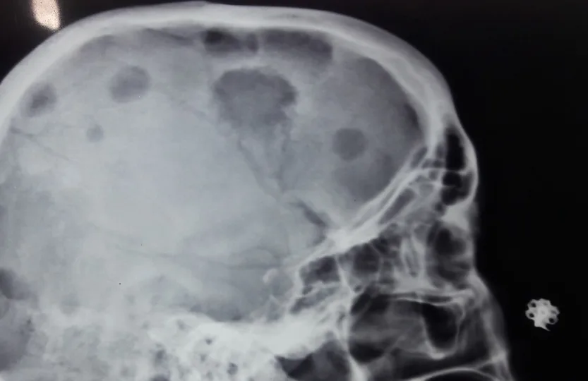
Lateral skull radiograph.
Image by Medicalpal, via Wikimedia Commons. Licence: CC BY-SA 4.0.
Protein studies
Laboratory results
Serum and urine protein studies are sent to characterise the raised globulin fraction.
| Parameter | Value | Reference |
|---|---|---|
| Serum electrophoresis | Dense monoclonal band, gamma regionRef No monoclonal band | No monoclonal band |
| Immunofixation | IgG kappa paraproteinRef No paraprotein | No paraprotein |
| Paraprotein concentration | 48 g/LRef Not detected | Not detected |
| Serum free light chain ratio (κ:λ) | 184Ref 0.26–1.65 | 0.26–1.65 |
| Urine Bence-Jones protein | PresentRef Absent | Absent |
| IgA / IgM | Both suppressedRef IgA 0.8–4.0, IgM 0.5–2.0 g/L | IgA 0.8–4.0, IgM 0.5–2.0 g/L |
Bone marrow biopsy
Pathology
Bone marrow aspirate and trephine show 42% plasma cells with atypical morphology, including binucleate forms. Flow cytometry confirms a kappa-restricted clonal population. Cytogenetics by FISH show t(4;14).