17 Sept 2026 · Case #012
Aortic dissection (Stanford type A)
This page works the case through in full, answer included. If you would rather reason it out yourself: Play this case
Diagnosis
Aortic dissection (Stanford type A)
First-line treatment
Emergency surgical repair, with IV beta blockade while awaiting theatre
How the diagnosis is reached
Pain that is maximal at the very first instant is the discriminator. Myocardial ischaemia builds over minutes as the muscle starves, whereas a tear happens all at once, and a tearing quality radiating to the back fits that mechanism. Everything else on examination follows from a flap propagating along the aorta. The inter-arm pressure difference and asymmetric pulses mean the flap has reached the brachiocephalic vessels and is intermittently obstructing them, and the early diastolic murmur is aortic regurgitation from a disrupted valve annulus, which puts the dissection in the ascending aorta. The mildly raised troponin is the trap this case is built around: a flap can shear a coronary ostium and produce genuine ischaemia, and anchoring on that number sends the patient to the catheter lab or, far worse, to thrombolysis, which is close to lethal here. The ECG is the counterweight, and it earns its place by what it lacks: sinus tachycardia with the tall voltages of left ventricular hypertrophy from years of untreated pressure, non-specific T-wave flattening in the lateral leads, and no ST elevation, no reciprocal depression and no territory that evolves across serial tracings. A troponin rise without any of that is not an occluded coronary artery. Long-standing untreated hypertension is the risk factor that made the wall vulnerable in the first place. A Stanford type A dissection involves the ascending aorta and is an operative emergency, with mortality climbing roughly 1 to 2 per cent per hour, so definitive treatment is emergency open repair. While theatre is readied, intravenous beta blockade with esmolol or labetalol brings heart rate under 60 and systolic pressure to 100 to 120, which lowers the shear force driving the tear onward. The order is not negotiable: beta blockade precedes any vasodilator, because a vasodilator given first provokes reflex tachycardia and extends the dissection. Uncomplicated type B dissections are managed medically.
Key clinical clues
- Pain maximal at onset, tearing, radiating between the shoulder blades
- Inter-arm blood pressure differential with an asymmetric radial pulse
- New early diastolic murmur of aortic regurgitation
- ECG without ST elevation despite a mildly raised troponin
- Widened mediastinum on chest radiograph, intimal flap on CT angiography
The case in full
Presenting complaint
Patient history
A 58-year-old man develops sudden, severe chest pain while gardening: a tearing sensation between the shoulder blades, at maximum intensity the instant it began. He has hypertension but stopped his medication a year ago.
Vital signs and examination
Vital signs
He is diaphoretic and restless, unable to find a comfortable position. The right radial pulse feels weaker than the left, and a soft early diastolic murmur is audible at the left sternal edge. Blood pressure is measured in both arms.
| Parameter | Value | Reference |
|---|---|---|
| Blood pressure (right arm) | 196/104 mmHg | – |
| Blood pressure (left arm) | 152/88 mmHg | – |
| Heart rate | 104 bpmRef 60–100 | 60–100 |
| Respiratory rate | 22 /minRef 12–20 | 12–20 |
| SpO₂ (room air) | 96 %Ref 95–100 | 95–100 |
Initial bloods
Laboratory results
Blood is drawn on arrival and the cardiology team is asked to review the results.
| Parameter | Value | Reference |
|---|---|---|
| High-sensitivity troponin T | 48 ng/LRef <14 | <14 |
| D-dimer | 6800 ng/mLRef <500 | <500 |
| Creatinine | 1.4 mg/dLRef 0.7–1.3 | 0.7–1.3 |
| Hemoglobin | 13.1 g/dLRef 13.5–17.5 | 13.5–17.5 |
| Lactate | 2.1 mmol/LRef 0.5–2.2 | 0.5–2.2 |
Electrocardiogram
ECG
A 12-lead ECG shows sinus tachycardia with left ventricular hypertrophy and non-specific T-wave flattening in the lateral leads. There is no ST elevation, no reciprocal depression, and no evolving territory on serial tracings.
Chest radiograph
Imaging
A chest radiograph is taken in the resuscitation bay.
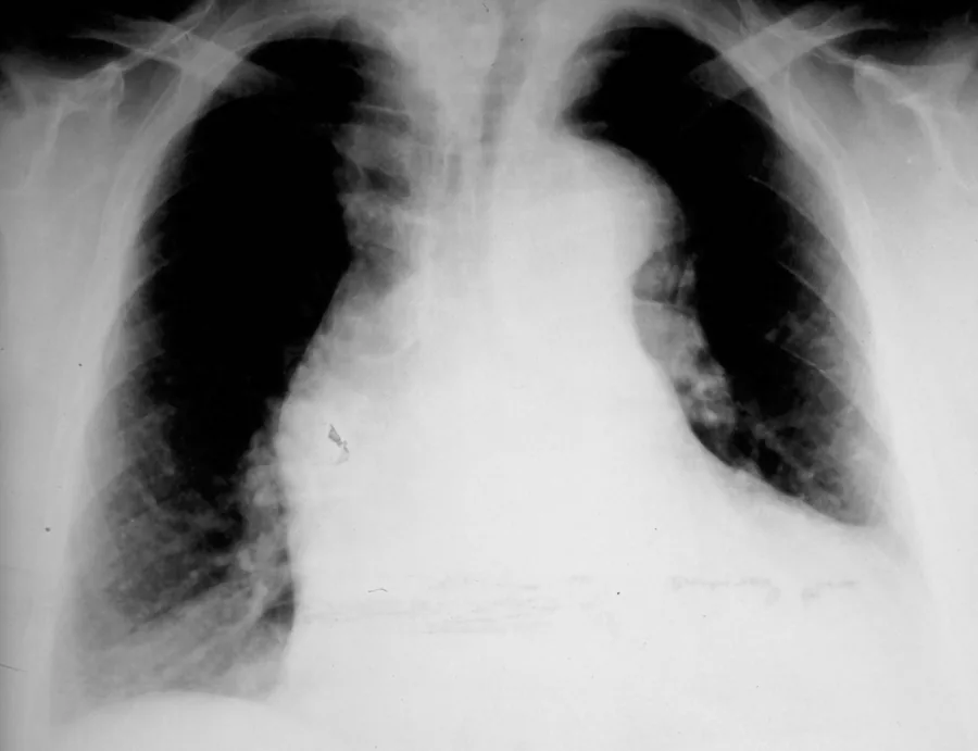
Frontal chest radiograph.
Image by J. Heuser, via Wikimedia Commons, with the numbered labels removed. Licence: CC BY-SA 3.0.
Cross-sectional imaging
Imaging
CT angiography shows an intimal flap 2 cm above the aortic valve, with true and false lumens running past the left subclavian into the descending aorta. There is a small pericardial effusion, and the ascending aorta is aneurysmal at 5.7 cm, predating today.