9 Sept 2026 · Case #004
ST-elevation myocardial infarction (STEMI)
This page works the case through in full, answer included. If you would rather reason it out yourself: Play this case
Diagnosis
ST-elevation myocardial infarction (STEMI)
First-line treatment
Primary percutaneous coronary intervention (PCI)
How the diagnosis is reached
Exertional chest pain with sweating and nausea is acute coronary syndrome until an ECG says otherwise, and here the ECG settles it. Three millimetres of ST elevation across V2 to V4 is a contiguous anterior territory rather than the diffuse saddle-shaped elevation of pericarditis, and the reciprocal ST depression in the inferior leads is what separates a true infarct from the mimics that also elevate ST segments; the absent friction rub argues the same way. Those leads also localise the culprit before angiography does, since V2 to V4 is left anterior descending territory. The troponin is the trap. Forty minutes in, mildly elevated is exactly right, since the rise lags the occlusion by hours, and a normal value would have excluded nothing. Nobody waits for it. Muscle downstream of the proximal LAD is dying now, so the only question left is which method reopens the artery soonest. That is primary PCI wherever it can be reached in time: mechanical reopening with a stent, target under 90 minutes from first contact. Past about 120, transfer delay costs more than the drug does, so a fibrinolytic (tenecteplase, alteplase) is given and the patient sent for angiography afterwards regardless. It is a fallback rather than an equal, reopening roughly half of arteries against over ninety per cent, with a bleeding risk PCI lacks. Either way aspirin and a P2Y12 inhibitor such as ticagrelor start immediately, since the clot is platelet-rich and an opened vessel re-occludes without them.
Key clinical clues
- Crushing chest pain radiating to arm/jaw with diaphoresis, classic ACS presentation
- ST elevation in V2–V4 localizing to the anterior wall (LAD territory)
- Anterior wall motion abnormality on echocardiogram
- Angiography confirming proximal LAD occlusion
- Time-critical reperfusion is the priority, PCI preferred, fibrinolysis if unavailable
The case in full
Presenting complaint
Patient history
A 58-year-old man presents to the emergency department with crushing central chest pain that started 40 minutes ago while shoveling snow. The pain radiates to his left arm and jaw and is associated with sweating and nausea.
Physical examination
Physical examination
He appears pale and diaphoretic. Cardiac and respiratory exams are otherwise unremarkable, with no murmurs or friction rub.
| Parameter | Value | Reference |
|---|---|---|
| Heart rate | 98 bpmRef 60–100 | 60–100 |
| Blood pressure | 152/94 mmHg | – |
| SpO₂ (room air) | 96 %Ref 95–100 | 95–100 |
Cardiac biomarkers
Laboratory results
A high-sensitivity troponin is drawn on arrival.
| Parameter | Value | Reference |
|---|---|---|
| High-sensitivity troponin | Mildly elevatedRef Below assay cutoff | Below assay cutoff |
12-lead ECG
ECG
The ECG shows 3 mm ST-segment elevation in leads V2 through V4, with reciprocal ST depression in the inferior leads.
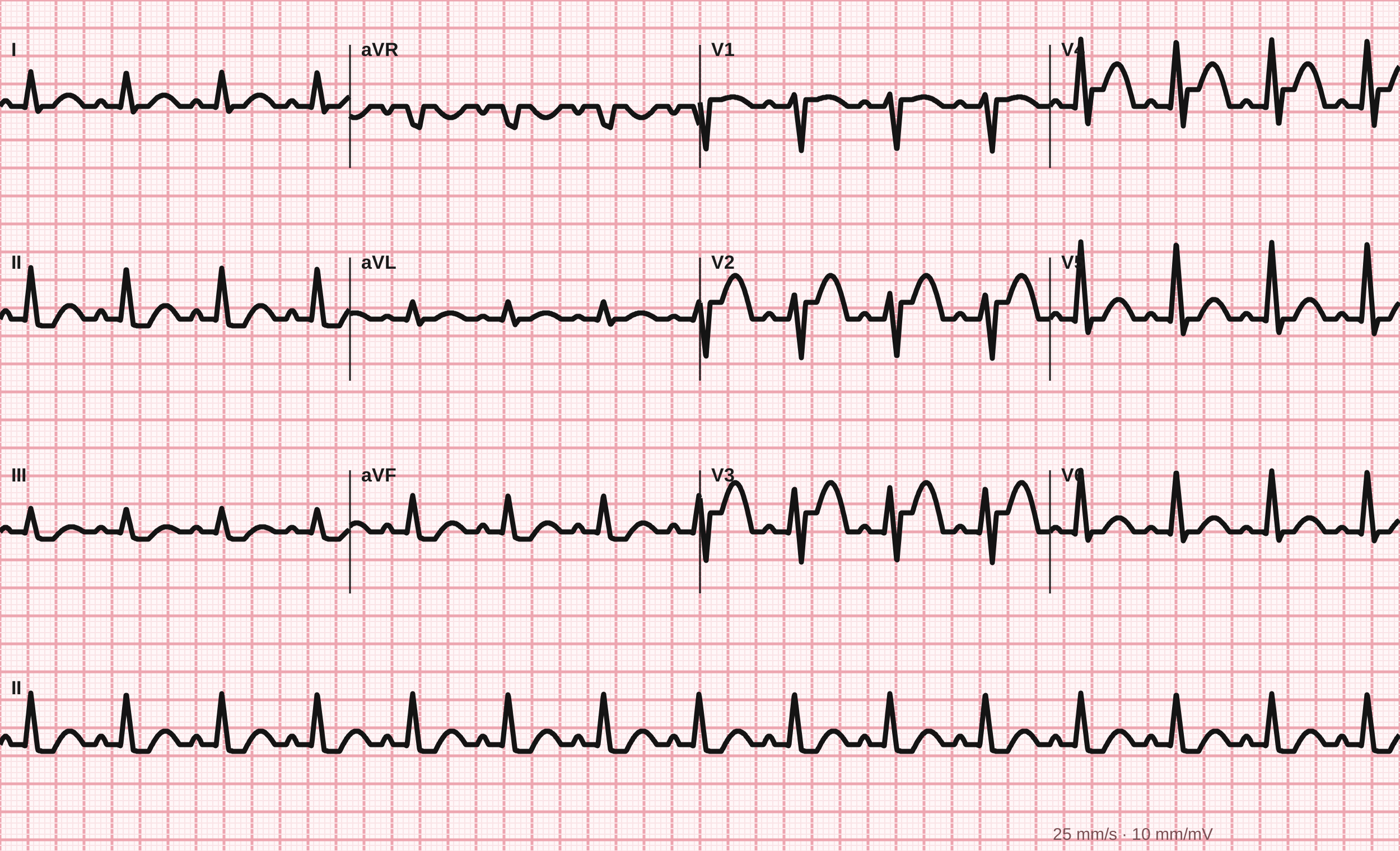
12-lead ECG, 25 mm/s.
Tracing drawn for MedGuesser, not a recording from a patient.
Bedside echocardiogram
Imaging
A focused echocardiogram shows a regional wall motion abnormality of the anterior wall with reduced overall ejection fraction.
Cardiac catheterization
Clinical course
The catheterization lab is activated. Coronary angiography reveals a complete occlusion of the proximal left anterior descending (LAD) artery.