10 Sept 2026 · Case #005
Pulmonary embolism (PE)
This page works the case through in full, answer included. If you would rather reason it out yourself: Play this case
Diagnosis
Pulmonary embolism (PE)
First-line treatment
Therapeutic anticoagulation
How the diagnosis is reached
Two risk factors compound here rather than merely coexist: fourteen hours of immobility three days ago, and an oestrogen-containing pill that raises clotting factor levels for as long as it is taken. Sudden pleuritic pain with hypoxia and a clear chest is the useful combination, because it is the absence of findings on auscultation that makes pneumonia and pneumothorax poor fits for this degree of hypoxia. The blood gas quietly confirms the mechanism. A pH of 7.49 with a PaCO2 of 29 is a respiratory alkalosis from blowing off carbon dioxide, meaning she is hyperventilating and still only managing 91 per cent, which is dead space rather than a ventilation problem. The ECG shows the same thing from another angle: sinus tachycardia, with a prominent S wave in lead I, a Q wave in lead III and an inverted T wave there, the S1Q3T3 pattern of an acutely strained right ventricle. It is worth recognising but not worth waiting for, specific enough to be a useful hint and far too insensitive to exclude anything, and plain sinus tachycardia is the commoner finding. She is haemodynamically stable, which is the finding that determines treatment. Anticoagulation alone is therefore enough, and a direct oral anticoagulant such as apixaban or rivaroxaban, both direct factor Xa inhibitors, is preferred for most patients; low molecular weight heparin is used instead in pregnancy and active cancer. It is started on suspicion rather than on the scan when suspicion is high, since the wait is the dangerous part. Systemic thrombolysis is held back for high-risk PE with haemodynamic instability, because the bleeding risk only justifies itself against an immediate threat to life.
Key clinical clues
- Risk factors: recent long-haul flight plus oral contraceptive use
- Sudden pleuritic chest pain, tachycardia, and hypoxia
- S1Q3T3 pattern on ECG
- CT pulmonary angiography showing bilateral filling defects
- DVT identified as the embolic source on lower extremity ultrasound
The case in full
Presenting complaint
Patient history
A 45-year-old woman presents with sudden-onset shortness of breath and right-sided pleuritic chest pain that began two hours ago. She returned from a 14-hour international flight three days ago and takes a combined oral contraceptive pill.
Physical examination
Physical examination
She is anxious and tachypneic but able to speak in full sentences. Breath sounds are equal bilaterally with no focal findings on auscultation.
| Parameter | Value | Reference |
|---|---|---|
| Heart rate | 118 bpmRef 60–100 | 60–100 |
| Respiratory rate | 26 /minRef 12–20 | 12–20 |
| SpO₂ (room air) | 91 %Ref 95–100 | 95–100 |
Laboratory studies
Laboratory results
D-dimer and an arterial blood gas are obtained.
| Parameter | Value | Reference |
|---|---|---|
| D-dimer | Markedly elevatedRef Below assay cutoff | Below assay cutoff |
| Arterial pH | 7.49Ref 7.35–7.45 | 7.35–7.45 |
| PaCO₂ | 29 mmHgRef 35–45 | 35–45 |
12-lead ECG
ECG
The ECG shows sinus tachycardia with an S1Q3T3 pattern, a prominent S wave in lead I, a Q wave in lead III, and an inverted T wave in lead III.
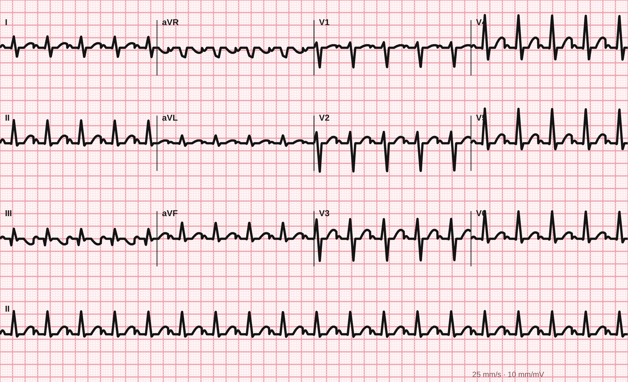
12-lead ECG, 25 mm/s.
Tracing drawn for MedGuesser, not a recording from a patient.
CT pulmonary angiography
Imaging
CT pulmonary angiography shows filling defects in the right and left main pulmonary arteries, consistent with bilateral emboli.
Lower extremity ultrasound
Imaging
Doppler ultrasound of the lower extremities reveals an occlusive thrombus in the right popliteal vein, identifying a likely embolic source.