8 Sept 2026 · Case #003
Classical Hodgkin lymphoma
This page works the case through in full, answer included. If you would rather reason it out yourself: Play this case
Diagnosis
Classical Hodgkin lymphoma
First-line treatment
ABVD chemotherapy
How the diagnosis is reached
A firm rubbery node that has grown painlessly for six weeks, with fever, drenching sweats and weight loss behind it, is lymphoma until a biopsy says otherwise; infection and reactive nodes do not behave this way. Two features then separate Hodgkin from the non-Hodgkin lymphomas before any pathology arrives. Alcohol-induced pain in the node is uncommon but carries real weight when present, and the disease has spread contiguously from neck to mediastinum rather than in the scattered skip lesions typical of non-Hodgkin disease. Hodgkin lymphoma travels station by station, which is why staging it is meaningful. The immunophenotype settles it, and the negatives do the work: CD30 and CD15 positive is consistent with Reed-Sternberg cells, but CD45 negative is the discriminator, since that leukocyte common antigen is expressed by nearly every other lymphoma. The eosinophilia and raised ESR reflect the cytokine-rich reactive background those cells recruit, which is most of the visible tumour. Treatment is ABVD, four drugs given together: doxorubicin, bleomycin, vinblastine and dacarbazine. Escalated BEACOPP controls high-risk advanced disease better but is markedly more toxic, and at 24 that trade matters, because cure already exceeds 80 per cent and he has decades in which infertility, secondary leukaemia, bleomycin lung injury and doxorubicin cardiotoxicity would be felt. Interim PET after two cycles is what allows the intensity to be raised or lowered on evidence rather than guessed at the start.
Key clinical clues
- Painless, rubbery cervical/supraclavicular lymphadenopathy with B symptoms in a young adult
- Alcohol-induced lymph node pain, an uncommon but fairly specific finding
- Elevated ESR/LDH with contiguous nodal spread into the mediastinum on imaging
- Biopsy showing large binucleated Reed-Sternberg ("owl's eye") cells in a mixed inflammatory background
- CD30+/CD15+, CD20-/CD45- immunophenotype confirming classical Hodgkin lymphoma
The case in full
Presenting complaint
Patient history
A 24-year-old man has had a painless, enlarging lump on the left side of his neck for six weeks, with recurrent fevers, drenching night sweats and 5 kg of weight loss over the past fortnight. The lump turns painful within minutes of drinking alcohol.
Physical examination
Physical examination
Examination reveals a firm, rubbery, non-tender lymph node measuring about 3 cm in the left supraclavicular region, with no overlying skin changes and no other palpable lymphadenopathy. The spleen tip is just palpable below the costal margin.
Blood tests
Laboratory results
Routine bloodwork is obtained alongside inflammatory markers.
| Parameter | Value | Reference |
|---|---|---|
| Hemoglobin | 10.8 g/dLRef 13.5–17.5 | 13.5–17.5 |
| ESR | 62 mm/hrRef 0–15 | 0–15 |
| LDH | 310 U/LRef 140–280 | 140–280 |
| Eosinophil count | 0.9 ×10⁹/LRef 0–0.5 | 0–0.5 |
Chest imaging
Imaging
Contrast-enhanced CT of the chest shows a bulky anterior mediastinal mass along with enlarged lymph nodes extending contiguously from the neck into the mediastinum (an orderly, contiguous pattern of spread rather than scattered skip lesions).
Lymph node biopsy: histology
Pathology
An excisional biopsy of the supraclavicular node is performed. The H&E section is shown below.
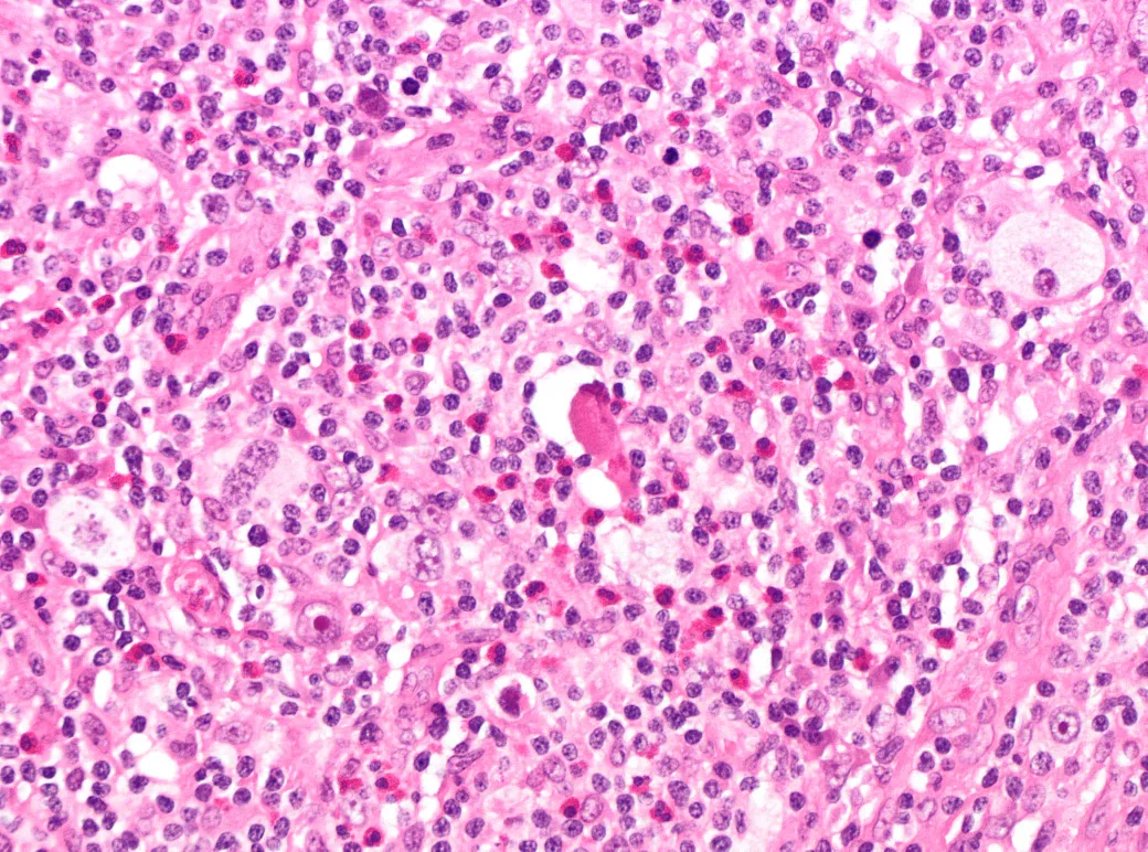
H&E stain, high power.
Image by Mark Ong, via Wikimedia Commons. Licence: CC BY-SA 4.0.
Immunohistochemistry
Clinical course
The large atypical cells stain positive for CD30 and CD15, and are negative for CD20 and CD45 (leukocyte common antigen), a pattern distinct from the small reactive lymphocytes surrounding them.