13 Oct 2026 · Case #038
Sarcoidosis
This page works the case through in full, answer included. If you would rather reason it out yourself: Play this case
Diagnosis
Sarcoidosis
First-line treatment
Oral corticosteroids
How the diagnosis is reached
Bilateral hilar lymphadenopathy, erythema nodosum, ankle arthritis and fever together are Löfgren syndrome, the acute face of sarcoidosis, and the combination is specific enough that biopsy is sometimes not needed at all. The work of the case is excluding the two conditions that produce hilar nodes and granulomas and are treated in the opposite way. Tuberculosis is the first: its granulomas are caseating, with cheesy necrosis at the centre, whereas these are well formed and necrosis-free, and the negative Ziehl–Neelsen, culture and interferon-gamma release assay close it off. That distinction is not academic, since the treatment for sarcoidosis is corticosteroids, which would be actively dangerous in untreated tuberculosis. Lymphoma is the second, and the symmetry argues against it: lymphoma tends to produce asymmetrical, bulky mediastinal disease rather than mirror-image hilar nodes with clear lungs. The remaining numbers fit the mechanism. Lymphopenia is characteristic, because T cells are sequestered into the affected tissue rather than depleted. The hypercalcaemia comes from granuloma macrophages expressing 1α-hydroxylase and converting vitamin D to its active form outside normal feedback control, which is also why these patients are told to avoid supplements and strong sun. Serum ACE is produced by the granulomas themselves; it is too insensitive and non-specific to diagnose on, but it fits. Löfgren syndrome carries the best prognosis of any sarcoid presentation, most cases remitting within two years, which is why much of sarcoidosis needs no treatment at all and non-steroidal anti-inflammatories would ordinarily cover the arthritis and the erythema nodosum. What changes the calculation here is the calcium of 2.78 mmol/L. Hypercalcaemia is a recognised indication to treat, alongside progressive lung disease, cardiac or neurological involvement and eye disease, so she gets oral prednisolone at moderate dose, tapered over six to twelve months, with methotrexate as the usual steroid-sparing second line.
Key clinical clues
- Bilateral hilar lymphadenopathy with clear lung fields on the radiograph
- Erythema nodosum with ankle arthritis and fever, the Löfgren combination
- Granulomas that are well formed and free of central necrosis, unlike tuberculosis
- Negative Ziehl–Neelsen, mycobacterial culture and interferon-gamma release assay
- Hypercalcaemia with lymphopenia and a raised ACE, from granuloma activity itself
The case in full
Presenting complaint
Patient history
A 29-year-old woman has had six weeks of dry cough, breathlessness climbing stairs and vague chest discomfort. She is tired and has had low-grade fevers.
Skin and joints
Symptoms
Three weeks ago tender red nodules appeared over both shins, and both ankles have been painful and swollen since. She has not travelled, has never smoked and takes no medication.
Examination and observations
Physical examination
There are raised, tender, dusky nodules over both shins and both ankles are swollen. The chest is clear on auscultation and no lymph nodes are palpable in the neck or axillae.
| Parameter | Value | Reference |
|---|---|---|
| Temperature | 37.6 °CRef 36.1–37.5 | 36.1–37.5 |
| SpO₂ (room air) | 96 %Ref 95–100 | 95–100 |
| Respiratory rate | 18 /minRef 12–20 | 12–20 |
| Heart rate | 84 bpmRef 60–100 | 60–100 |
Blood tests
Laboratory results
Routine bloods and a metabolic screen are sent.
| Parameter | Value | Reference |
|---|---|---|
| White cell count | 6.8 ×10⁹/LRef 4.0–11.0 | 4.0–11.0 |
| Lymphocytes | 0.9 ×10⁹/LRef 1.0–3.0 | 1.0–3.0 |
| CRP | 32 mg/LRef <5 | <5 |
| ESR | 54 mm/hRef <20 | <20 |
| Corrected calcium | 2.78 mmol/LRef 2.20–2.60 | 2.20–2.60 |
| Serum ACE | 96 U/LRef 8–52 | 8–52 |
| Creatinine | 71 µmol/LRef 45–90 | 45–90 |
Chest imaging
Imaging
Her chest radiograph is shown below. CT then confirms bilateral hilar and mediastinal lymphadenopathy with a few small nodules along the bronchovascular bundles, and no cavitation, mass or effusion.
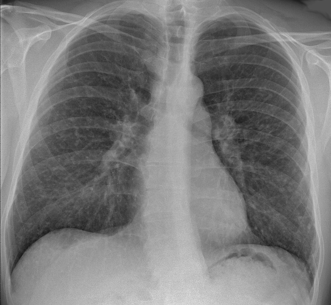
Chest radiograph, PA.
Image by Mikael Häggström, via Wikimedia Commons. Licence: CC0.
Node biopsy
Pathology
Endobronchial ultrasound-guided biopsy of a hilar node shows well-formed granulomas with no necrosis at their centres. Ziehl–Neelsen staining, mycobacterial culture and fungal stains are all negative, as is an interferon-gamma release assay.