14 Oct 2026 · Case #039
Rhabdomyolysis
This page works the case through in full, answer included. If you would rather reason it out yourself: Play this case
Diagnosis
Rhabdomyolysis
First-line treatment
Aggressive IV fluid resuscitation
How the diagnosis is reached
The whole case turns on one line of the urinalysis. The dipstick is strongly positive for blood, yet microscopy finds no red cells, and that combination has a single explanation: the dipstick detects haem, so it reads positive for myoglobin exactly as it does for haemoglobin. What is colouring this urine is muscle pigment, not bleeding. That immediately separates it from the glomerular causes of dark urine, where dysmorphic red cells and red cell casts are the finding. A creatine kinase of 84,000 confirms the source, and levels above roughly 5,000 mark the point where kidney injury becomes likely. The mechanism of the injury is threefold: myoglobin casts obstruct the tubules, haem is directly toxic to tubular cells, and fluid sequestered into swollen muscle leaves the patient intravascularly dry, driving renal vasoconstriction. The electrolytes are simply the contents of broken muscle cells appearing in the blood, which is why potassium and phosphate rise together while calcium falls as it deposits in damaged muscle. The raised AST is muscle in origin rather than liver, a common source of confusion. The precipitant here is unaccustomed exertion in someone deconditioned and dehydrated, the classic exertional pattern, and the ECG is the reminder that the potassium, not the kidney, is what can kill him in the next hour: the T waves across the praecordial leads are tall, narrow and symmetrically peaked on a QRS that is still narrow, the earliest change of hyperkalaemia and the one that precedes the widening QRS and sine wave pattern that follow if it is left alone. So the potassium is treated first and in parallel: calcium gluconate to stabilise the myocardium, insulin with dextrose to drive potassium back into cells. The kidney is then saved with volume. Early, large-volume crystalloid is the only intervention shown to prevent the injury, flushing myoglobin through the tubules before it can obstruct them, titrated to a urine output of 200 to 300 mL per hour, which often means several litres on the first day. Hartmann's and other potassium-containing fluids are avoided while the potassium is high. Bicarbonate and mannitol are traditional additions on weak evidence, and dialysis is held back for refractory hyperkalaemia, acidosis or anuria.
Key clinical clues
- Cola-coloured urine and severe muscle pain after an unaccustomed heavy workout
- Dipstick strongly positive for blood but no red cells on microscopy, so myoglobin rather than bleeding
- Creatine kinase of 84,000, far above the threshold at which kidney injury becomes likely
- Hyperkalaemia and hyperphosphataemia with a low calcium, the signature of muscle breakdown
- Peaked T waves on the ECG, making the potassium the immediate threat
The case in full
Presenting complaint
Patient history
A 24-year-old man has had severe aching in both thighs and shoulders for two days, and today noticed that his urine has turned the colour of cola. He can barely climb stairs.
Background
Patient history
He returned to the gym after months away and did an unusually heavy leg and shoulder session three days ago. He takes no regular medication, but has been using a pre-workout supplement and drinking very little water.
Examination and observations
Vital signs
Both thighs and shoulders are diffusely tender and feel firm, with pain on any movement. There is no rash and no joint swelling. He has passed very little urine since yesterday.
| Parameter | Value | Reference |
|---|---|---|
| Temperature | 37.2 °CRef 36.1–37.5 | 36.1–37.5 |
| Heart rate | 96 bpmRef 60–100 | 60–100 |
| Blood pressure | 128/78 mmHg | – |
| Respiratory rate | 16 /minRef 12–20 | 12–20 |
Urinalysis
Laboratory results
A fresh urine sample is dipped and then examined under the microscope.
| Parameter | Value | Reference |
|---|---|---|
| Appearance | Dark brownRef Straw | Straw |
| Dipstick blood | 3+Ref Negative | Negative |
| Microscopy | No red cells seenRef No red cells | No red cells |
| Dipstick protein | 1+Ref Negative | Negative |
| Casts | Pigmented granular castsRef None | None |
Blood tests
Laboratory results
Bloods are sent urgently.
| Parameter | Value | Reference |
|---|---|---|
| Creatine kinase | 84,000 U/LRef <200 | <200 |
| Creatinine | 268 µmol/LRef 60–110 | 60–110 |
| Potassium | 6.1 mmol/LRef 3.5–5.0 | 3.5–5.0 |
| Corrected calcium | 1.92 mmol/LRef 2.20–2.60 | 2.20–2.60 |
| Phosphate | 2.4 mmol/LRef 0.8–1.5 | 0.8–1.5 |
| Bicarbonate | 17 mmol/LRef 22–26 | 22–26 |
| AST | 420 U/LRef <40 | <40 |
Bedside monitoring
ECG
The ECG shows tall, peaked T waves across the praecordial leads. There was no trauma, no seizure and no long period spent lying immobile, and an autoimmune myositis screen is negative.
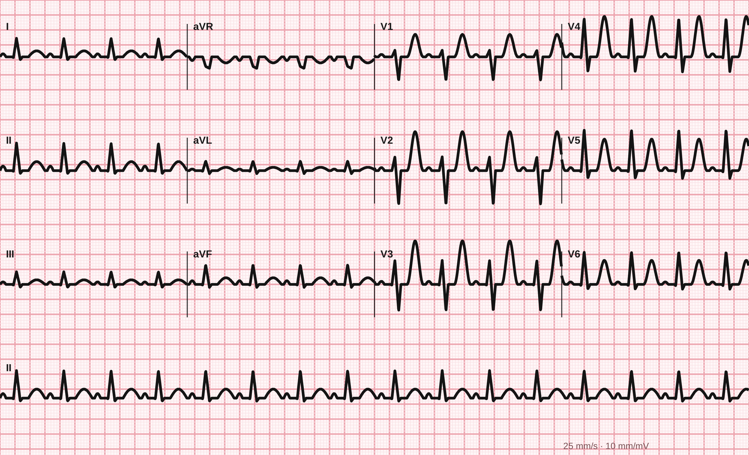
12-lead ECG, 25 mm/s.
Tracing drawn for MedGuesser, not a recording from a patient.