21 Oct 2026 · Case #046
Idiopathic pulmonary fibrosis (IPF)
This page works the case through in full, answer included. If you would rather reason it out yourself: Play this case
Diagnosis
Idiopathic pulmonary fibrosis (IPF)
First-line treatment
Antifibrotic therapy (pirfenidone or nintedanib)
How the diagnosis is reached
Crackles that coughing does not clear, clubbing and a year and a half of breathlessness put this in the interstitium rather than the airways, and the spirometry settles the compartment before the scan names the disease. A low FVC with an FEV1/FVC ratio of 0.83 is restriction, the opposite of the obstruction twenty-five pack-years invites you to expect, and a gas transfer of 44 per cent, down further than the volumes are, is exchange failing across a thickened membrane. The CT then does the work. Honeycombing means clustered cysts of similar size stacked in rows directly under the pleura, and with basal and posterior predominance, traction bronchiectasis and architectural distortion, that is the usual interstitial pneumonia pattern. Each absent finding removes a named alternative. No perilymphatic nodularity and no hilar nodes, so not sarcoidosis. No mosaic attenuation or air trapping, so not hypersensitivity pneumonitis, which the negative precipitins argue against as well. No pleural plaque, so not asbestosis, which his working life never exposed him to. No ground-glass, which is what a cellular non-specific interstitial pneumonia would show. Negative ANA, rheumatoid factor, anti-CCP, ANCA and myositis antibodies alongside no synovitis, rash or Raynaud remove connective tissue disease, and a normal BNP with normal ventricular function removes the heart. Idiopathic is what remains, arrived at deliberately rather than by default. Treatment does not reverse fibrosis and is not meant to. An antifibrotic, pirfenidone or nintedanib, roughly halves the annual rate of FVC decline, and his own plot is the argument for starting now, since he has lost half a litre in eighteen months. The two are comparable in effect and differ in what they cost: pirfenidone photosensitivity and nausea, nintedanib diarrhoea and raised transaminases, so the choice follows tolerance. Corticosteroids are the trap: they help the inflammatory interstitial diseases this has just been separated from, and in IPF they increased mortality when they were trialled. Alongside the drug go ambulatory oxygen for his desaturation, pulmonary rehabilitation, and a transplant referral now, because at 68 that door is closing.
Key clinical clues
- Eighteen months of exertional breathlessness with a dry unproductive cough
- Fine end-inspiratory crackles that coughing does not clear, and clubbed fingers
- Restriction with a preserved FEV1/FVC ratio, and gas transfer down to 44 per cent
- Half a litre of vital capacity lost across four clinic visits
- Subpleural honeycombing worst at the bases, with no ground-glass, nodules or pleural plaque
The case in full
Presenting complaint
Patient history
A 68-year-old man has been getting steadily more breathless for eighteen months and can no longer manage one flight of stairs without stopping halfway. He has a dry cough that nothing he has tried touches. There is no wheeze, no sputum, no chest pain and no weight loss.
Background
Patient history
He smoked twenty-five pack-years and stopped fifteen years ago. He worked as an accountant: no mining, no farming, no shipyard, no stonework. He keeps no birds, has no visible damp at home and does not use a hot tub. He takes ramipril alone, and has never taken amiodarone, methotrexate or nitrofurantoin. He has no joint pain, no rash, no dry eyes and no colour change in his fingers in the cold.
Examination and observations
Physical examination
There are fine crackles at both lung bases through the whole of inspiration, and they do not clear when he coughs. The fingers are clubbed. The chest is otherwise quiet with no wheeze, the jugular venous pressure is normal and there is no ankle swelling. There is no rash, no synovitis and no tightening of the skin over the fingers.
| Parameter | Value | Reference |
|---|---|---|
| Oxygen saturation, resting | 94 %Ref ≥95 | ≥95 |
| Oxygen saturation after 40 m walk | 86 %Ref ≥95 | ≥95 |
| Respiratory rate | 20 /minRef 12–20 | 12–20 |
| Heart rate | 88 bpmRef 60–100 | 60–100 |
| Temperature | 36.5 °CRef 36.1–37.5 | 36.1–37.5 |
Lung function
Laboratory results
Full pulmonary function testing is done, and the vital capacities recorded at his clinic visits over the last eighteen months are plotted alongside.
| Parameter | Value | Reference |
|---|---|---|
| Forced vital capacity (FVC) | 2.31 (68% predicted) LRef >80% predicted | >80% predicted |
| FEV1 | 1.92 (71% predicted) LRef >80% predicted | >80% predicted |
| FEV1/FVC ratio | 0.83Ref >0.70 | >0.70 |
| Total lung capacity | 71% predictedRef 80–120% predicted | 80–120% predicted |
| DLCO (gas transfer) | 44% predictedRef >80% predicted | >80% predicted |
Ruling out the mimics
Laboratory results
The secondary causes of a fibrotic lung are screened for before the scan is reported.
| Parameter | Value | Reference |
|---|---|---|
| Antinuclear antibody | NegativeRef Negative | Negative |
| Rheumatoid factor and anti-CCP | Both negativeRef Negative | Negative |
| ANCA | NegativeRef Negative | Negative |
| Myositis antibody panel | NegativeRef Negative | Negative |
| Avian and fungal precipitins | NegativeRef Negative | Negative |
| BNP | 38 ng/LRef <100 | <100 |
| Echocardiogram | Normal ventricular function, no valve lesionRef Normal | Normal |
High-resolution CT
Imaging
A high-resolution CT of the chest is performed without contrast. An axial slice through the lower zones is shown below. There is coarse reticulation with clustered cystic spaces of similar size stacked in rows immediately beneath the pleura, worst posteriorly and at both bases, with distortion of the architecture and dilated airways running out into the fibrosis. There is no ground-glass, no mosaic attenuation, no nodularity, no enlarged hilar node and no pleural plaque or effusion. The upper zones are comparatively spared.
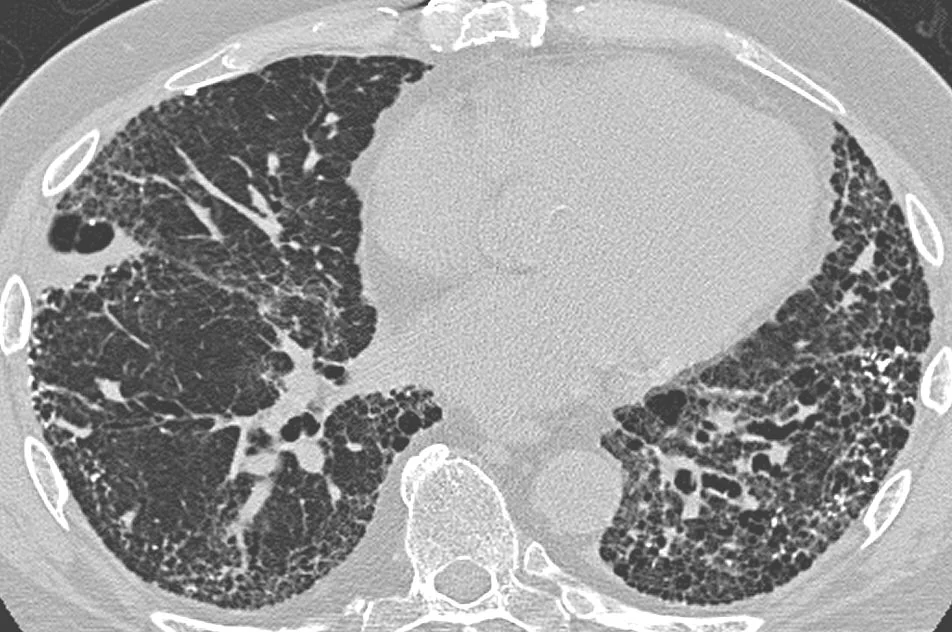
HRCT chest, axial.
Image by Yale Rosen, contributed by Darel Heitkamp, via Wikimedia Commons. Licence: CC BY-SA 2.0.