19 Oct 2026 · Case #044
Complete hydatidiform mole (molar pregnancy)
This page works the case through in full, answer included. If you would rather reason it out yourself: Play this case
Diagnosis
Complete hydatidiform mole (molar pregnancy)
First-line treatment
Suction curettage
How the diagnosis is reached
A fundus at the umbilicus at ten weeks is the discriminator, because the other causes of first-trimester bleeding leave the uterus small for dates or unchanged: a missed or threatened miscarriage, an ectopic, a cervical lesion. Large for dates with no audible heartbeat leaves two candidates, a multiple pregnancy or trophoblast growing without a fetus, and the scan separates them. What fills the cavity is echogenic tissue studded with small cystic spaces, hydropic villi seen one by one, with no sac, no pole and no heart beating anywhere in it; a twin pregnancy at this size would show two of everything instead. The hCG of 312 000 is roughly three times what a normal ten-week pregnancy peaks at, and it accounts for findings that would otherwise read as a second illness. Vomiting severe enough to cost 4 kg tracks hCG. So does the thyroid picture: hCG resembles TSH closely enough that a concentration this high stimulates the TSH receptor directly, which is why the free T4 is raised, the TSH suppressed and the receptor antibodies negative, where Graves disease would have them positive. The bulky ovaries are theca lutein cysts driven by the same hormone, and they regress as it falls, so they are left alone. Treatment is suction curettage, and the technique carries the reasoning: a molar uterus is soft, large and highly vascular, so it is emptied by suction under ultrasound guidance, with an oxytocin infusion held back until the cavity is nearly empty, because squeezing a vascular uterus around trophoblast risks pushing it into the circulation. Medical evacuation with a prostaglandin is avoided for the same reason. She is rhesus negative, so anti-D is given, and the tissue goes for histology and ploidy. Surveillance matters as much as the operation: serial hCG until it is undetectable, since roughly one mole in six persists as gestational trophoblastic neoplasia, and reliable contraception while it is watched, because a new pregnancy would hide the very rise being looked for.
Key clinical clues
- Ten weeks by dates, with the fundus already up at the umbilicus
- Eight days of dark bleeding with pale fragments in it, and no pain at all
- hCG of 312 000, about three times a normal ten-week peak
- Thyrotoxic biochemistry with negative TSH receptor antibodies
- A cavity full of small cystic spaces with no fetal pole and no fetal heart
The case in full
Presenting complaint
Patient history
A 24-year-old woman is ten weeks by her last period and has had dark brown vaginal bleeding on and off for eight days, twice with small pale fragments in it. She has vomited most days for three weeks, cannot keep breakfast down and has lost 4 kg. She has no abdominal pain.
Obstetric history
Patient history
This is her second pregnancy. The first ended in an uncomplicated delivery two years ago with no sickness worth mentioning. A home test was positive at six weeks, she has had no antenatal appointment yet, and she takes only folic acid. She has never had thyroid trouble and nobody in the family has.
Examination and observations
Physical examination
The uterus is palpable at the level of the umbilicus, far larger than ten weeks should be, and soft. No fetal heart is audible on a handheld Doppler. The cervix is closed with dark blood in the vault, and both adnexa feel bulky but not tender. Her hands are warm and sweaty and there is a fine tremor of the outstretched fingers.
| Parameter | Value | Reference |
|---|---|---|
| Fundal height | At the umbilicusRef Not palpable before 12 weeks | Not palpable before 12 weeks |
| Heart rate | 112 bpmRef 60–100 | 60–100 |
| Blood pressure | 124/76 mmHg | – |
| Temperature | 36.9 °CRef 36.1–37.5 | 36.1–37.5 |
| Fetal heart on Doppler | Not audibleRef Audible from 12 weeks | Audible from 12 weeks |
Blood tests
Laboratory results
Blood is taken in the early pregnancy unit.
| Parameter | Value | Reference |
|---|---|---|
| Serum beta-hCG | 312 000 IU/LRef Up to 100 000 at 10 weeks | Up to 100 000 at 10 weeks |
| Haemoglobin | 98 g/LRef 115–165 | 115–165 |
| Platelets | 268 ×10⁹/LRef 150–400 | 150–400 |
| Creatinine | 58 µmol/LRef 45–90 | 45–90 |
| ALT | 26 U/LRef <35 | <35 |
| Blood group | A rhesus negativeRef Not applicable | Not applicable |
Narrowing it down
Laboratory results
The thyroid function is checked because of the tremor, and the other explanations for her presentation are screened for.
| Parameter | Value | Reference |
|---|---|---|
| TSH | <0.01 mU/LRef 0.4–4.0 | 0.4–4.0 |
| Free T4 | 27 pmol/LRef 9–19 | 9–19 |
| TSH receptor antibodies | NegativeRef Negative | Negative |
| Urine protein | NegativeRef Negative | Negative |
| Chest radiograph | ClearRef Clear | Clear |
| Coagulation screen | NormalRef Normal | Normal |
Pelvic ultrasound
Imaging
A transvaginal scan is performed and the image is shown below. The cavity is filled with echogenic tissue carrying numerous small anechoic spaces of varying size. No gestational sac, yolk sac, fetal pole or fetal heart can be found anywhere within it. Both ovaries measure over 6 cm and hold multiple thin-walled cysts. There is no free fluid.
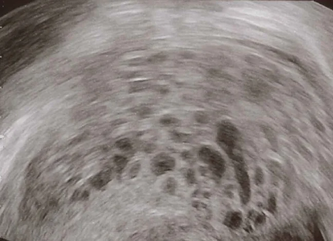
Transvaginal pelvic ultrasound.
Image by Mikael Häggström, via Wikimedia Commons, cropped to remove the scanner's own annotations. Licence: CC0.