18 Oct 2026 · Case #043
Autosomal dominant polycystic kidney disease (ADPKD)
This page works the case through in full, answer included. If you would rather reason it out yourself: Play this case
Diagnosis
Autosomal dominant polycystic kidney disease (ADPKD)
First-line treatment
Tolvaptan and blood pressure control
How the diagnosis is reached
Two ballotable flank masses is the finding that closes the differential, because the things that enlarge one kidney do not enlarge both: a renal cell carcinoma, an obstructed system, a single large simple cyst. The CT then names the architecture. Cysts of every size through both kidneys, with hepatic cysts beside them, is where the two conditions that could otherwise mimic this fall away, since neither the few cysts that accumulate with age nor the acquired cystic disease of long dialysis involves the liver or reaches this size in a man of 38. The pedigree finishes it: a father on dialysis at 52 who died of an intracranial bleed and an affected aunt is disease in two successive generations, which is dominant inheritance rather than the recessive form, and the recessive form declares itself in infancy with oligohydramnios rather than in the fourth decade with flank ache. The haemoglobin is the quiet finding worth cashing in. At an eGFR of 46 most chronic kidney disease is already anaemic, because a scarred interstitium makes less erythropoietin; here the cyst epithelium goes on producing it, so a normal haemoglobin at this stage is characteristic rather than reassuring. The visible blood was a cyst bleeding into itself and settles on its own, and a trace of protein is far too little for glomerular disease. Treatment has two halves and the first is the one that changes his outcome: blood pressure to target, and with an ACE inhibitor or an angiotensin receptor blocker rather than the dihydropyridine calcium channel blocker he is on, since renin-angiotensin blockade slows the decline in this disease specifically. Tolvaptan, a vasopressin V2 receptor antagonist, is the disease-modifying agent and slows both cyst growth and the fall in eGFR, but it is reserved for demonstrated rapid progression: it causes enormous thirst and urine volumes, and it needs monthly liver monitoring at first because it can cause serious hepatotoxicity. The 4 mm aneurysm is watched rather than repaired, and controlling his pressure is what protects it.
Key clinical clues
- Hypertension from the age of 33, treated but still not at target
- Two days of painless visible haematuria that settled without treatment
- Two affected generations: father on dialysis at 52, then dead of an intracranial bleed
- Bilateral ballotable flank masses, with cysts in the liver as well as both kidneys
- A normal haemoglobin despite an eGFR of 46
The case in full
Presenting complaint
Patient history
A 38-year-old man has had a dull ache low in both flanks for about six months, worse after a long stretch at his desk and never relieved by anything he has tried. Last week his urine was frankly red for two days and then cleared on its own. He has passed no stones, and had no fever and no burning.
Background
Patient history
He was told his blood pressure was high at 33 and has taken amlodipine since. His father began dialysis at 52 and died six years later of a bleed in the head; a paternal aunt is on the transplant list. He takes nothing else, has never smoked, and drinks a few beers at the weekend.
Examination and observations
Physical examination
He looks entirely well. Both flanks are full, and on each side a large irregular mass can be balloted between the hands and moves down on inspiration. There is no bruit over either renal angle, no ankle swelling and no rash.
| Parameter | Value | Reference |
|---|---|---|
| Blood pressure | 158/98 mmHgRef <140/90 | <140/90 |
| Heart rate | 76 bpmRef 60–100 | 60–100 |
| Temperature | 36.7 °CRef 36.1–37.5 | 36.1–37.5 |
| Respiratory rate | 14 /minRef 12–20 | 12–20 |
Blood and urine tests
Laboratory results
Bloods and a urine sample are sent from clinic.
| Parameter | Value | Reference |
|---|---|---|
| Creatinine | 152 µmol/LRef 60–110 | 60–110 |
| Estimated GFR | 46 mL/min/1.73m²Ref >90 | >90 |
| Urea | 9.8 mmol/LRef 2.5–7.8 | 2.5–7.8 |
| Haemoglobin | 148 g/LRef 130–170 | 130–170 |
| Potassium | 4.4 mmol/LRef 3.5–5.0 | 3.5–5.0 |
| Urate | 0.48 mmol/LRef 0.20–0.42 | 0.20–0.42 |
| Urine dipstick | Blood 1+, protein traceRef Negative | Negative |
| Urine albumin:creatinine ratio | 9 mg/mmolRef <3 | <3 |
Cross-sectional imaging
Imaging
A contrast-enhanced CT of the abdomen is arranged after the episode of visible blood. The coronal reconstruction is shown below. Both renal outlines are grossly enlarged and almost entirely replaced by rounded fluid-filled spaces of widely varying size, a few of them denser than the rest. Several similar lesions sit in the right lobe of the liver. There is no hydronephrosis, no calculus and no solid enhancing mass.
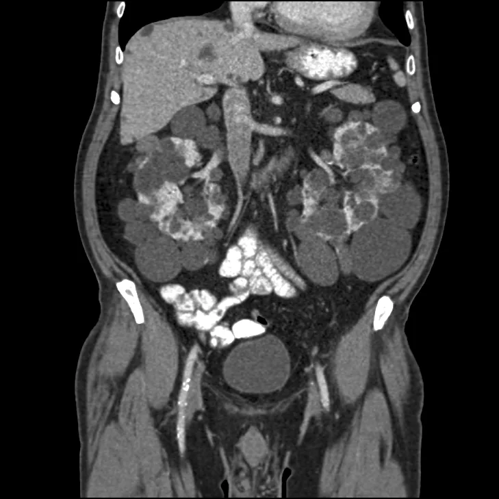
Abdominal CT, coronal reconstruction.
Image by Steven Fruitsmaak, via Wikimedia Commons. Licence: CC BY-SA 3.0.
Further assessment
Clinical course
Because of the family history, MR angiography of the intracranial vessels is performed and shows a 4 mm saccular aneurysm at the anterior communicating artery. Echocardiography shows mitral valve prolapse with trivial regurgitation and a normally sized left ventricle with a thickened wall. There is no diverticular disease on the abdominal scan.