22 Oct 2026 · Case #047
Gout
This page works the case through in full, answer included. If you would rather reason it out yourself: Play this case
Diagnosis
Gout
First-line treatment
Colchicine or an NSAID
How the diagnosis is reached
A hot swollen knee with a temperature of 38.1, a CRP of 148 and 42 000 white cells per microlitre of joint fluid is septic arthritis until the aspirate says otherwise, which is why the joint is tapped before the first antibiotic dose: a dose given first can sterilise the culture and leave the question open. The crystals answer it. Needles that turn yellow along the compensator axis and blue across it are negatively birefringent: monosodium urate. Calcium pyrophosphate, the other crystal that inflames a knee at this age, is the mirror image, rhomboid and positive where this is needle-shaped and negative, so one field separates gout from pseudogout. Crystals inside neutrophils rather than merely beside them is what makes them the cause rather than an old deposit. Two findings mislead at face value. The fever and neutrophilia belong to the attack: crystal-driven inflammation is a real innate immune response and runs systemic, so neither argues for infection, and the negative Gram stain with sterile fluid and blood closes that door. The urate of 0.38 is the more dangerous trap, because it is squarely normal, and urate commonly drops into the normal range during an acute attack; a normal level excludes nothing, and the value worth acting on is measured weeks later. The rest of the history explains why the disease arrived: a thiazide started four months ago cuts urate excretion, beer supplies purine and ethanol together, 12 kg adds insulin resistance, and stage 3 kidney disease removes the main route of clearance. The ear nodule is a tophus, which dates the problem well before tonight. For the attack, colchicine or an NSAID, and here colchicine, because an eGFR of 48 makes an NSAID a poor bargain; colchicine itself needs a reduced dose in renal impairment. An intra-articular or oral corticosteroid is the answer when neither drug is safe. Allopurinol is the definitive treatment and the wrong drug tonight: urate-lowering starts once the joint is quiet, introduced slowly under colchicine cover, because dropping urate abruptly sets off fresh attacks. The thiazide should be exchanged for something else.
Key clinical clues
- A monoarthritis of the knee at four in the morning, after two past attacks in the big toe
- Thiazide started four months ago, heavy beer intake, and stage 3 kidney disease
- Fever and a CRP of 148 with 42 000 synovial white cells, but a sterile stain and culture
- A firm painless nodule in the rim of the ear
- Needle crystals inside neutrophils, yellow along the compensator axis and blue across it
The case in full
Presenting complaint
Patient history
A 54-year-old man woke at four in the morning with his right knee hot, swollen and too painful to straighten. Twelve hours later he cannot bear weight on it. He has had two similar attacks over three years in the base of his right big toe, each settling within a week with nothing but paracetamol.
Background
Patient history
A thiazide diuretic was added for his blood pressure four months ago. He drinks six to eight beers most evenings and has put on 12 kg in two years. His family doctor has recorded stage 3 chronic kidney disease. The knee has had no injection and no injury, the skin over it has never been broken, and he reports no diarrhoea, no urethral discharge and no new sexual partner.
Examination and observations
Physical examination
The right knee is swollen and shiny with red skin over the patella, hot to the touch, held at twenty degrees of flexion and painful through any movement. A large effusion is ballottable. There is no breach in the skin, no tracking lymphangitis and no rash. Both first metatarsophalangeal joints are knobbly, and a firm painless nodule sits in the rim of the left ear.
| Parameter | Value | Reference |
|---|---|---|
| Temperature | 38.1 °CRef 36.1–37.5 | 36.1–37.5 |
| Heart rate | 96 bpmRef 60–100 | 60–100 |
| Blood pressure | 148/90 mmHgRef <140/90 | <140/90 |
| Respiratory rate | 16 /minRef 12–20 | 12–20 |
Blood tests
Laboratory results
Bloods and cultures are taken in the emergency department.
| Parameter | Value | Reference |
|---|---|---|
| CRP | 148 mg/LRef <5 | <5 |
| White cell count | 14.2 ×10⁹/LRef 4.0–11.0 | 4.0–11.0 |
| Neutrophils | 11.4 ×10⁹/LRef 2.0–7.5 | 2.0–7.5 |
| ESR | 74 mm/hRef <20 | <20 |
| Serum urate | 0.38 mmol/LRef 0.20–0.42 | 0.20–0.42 |
| Creatinine | 138 µmol/LRef 60–110 | 60–110 |
| Estimated GFR | 48 mL/min/1.73m²Ref >90 | >90 |
| Blood cultures | No growth at 48 hRef No growth | No growth |
Joint aspiration
Laboratory results
The knee is aspirated at the bedside before any antibiotic is given, and 40 mL of fluid is withdrawn.
| Parameter | Value | Reference |
|---|---|---|
| Appearance | Turbid, yellowRef Clear, straw-coloured | Clear, straw-coloured |
| Synovial white cell count | 42 000 /µLRef <200 | <200 |
| Synovial neutrophils | 88 %Ref <25 | <25 |
| Gram stain | No organisms seenRef No organisms | No organisms |
| Synovial fluid culture | No growth at 48 hRef No growth | No growth |
Synovial fluid microscopy
Pathology
A drop of the aspirate is examined under polarised light with a red compensator in the beam. The field is shown below. Needle-shaped crystals lie both free in the fluid and inside neutrophils. Each appears yellow where its long axis runs parallel to the axis of the compensator and blue where it runs across it.
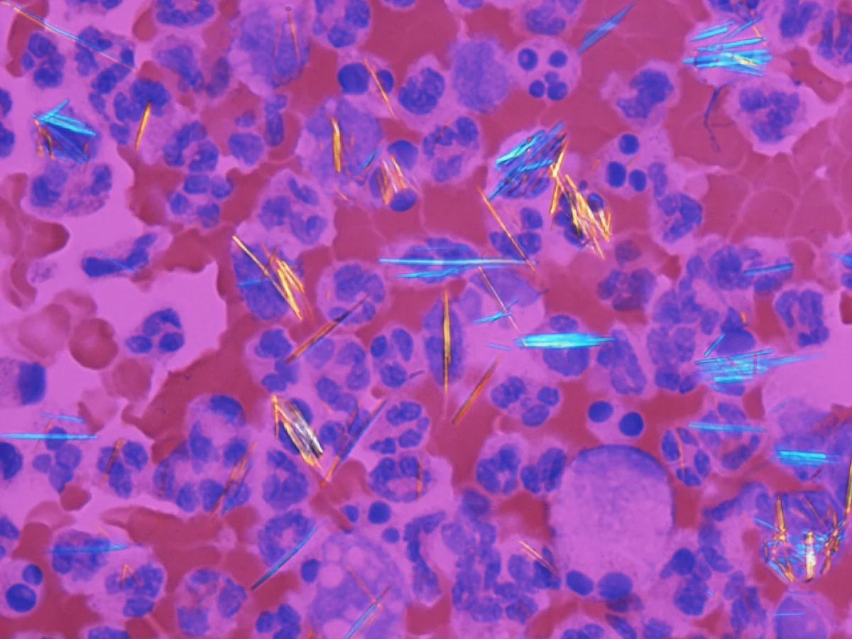
Synovial fluid, compensated polarised light.
Image by Gabriel Caponetti, via Wikimedia Commons. Licence: CC BY-SA 3.0.