23 Oct 2026 · Case #048
Central retinal artery occlusion (CRAO)
This page works the case through in full, answer included. If you would rather reason it out yourself: Play this case
Diagnosis
Central retinal artery occlusion (CRAO)
First-line treatment
Immediate referral on the acute stroke pathway
How the diagnosis is reached
The pupil finding is what narrows this before the retina is ever seen. A pupil that dilates when light is swung onto it means the afferent pathway of that eye is damaged, and that excludes everything in the media: a dense cataract, a vitreous haemorrhage and angle closure all cloud the picture without producing it, and the deep quiet anterior chamber with a pressure of 15 disposes of the last of those anyway. The fundus then names the vessel. A retina starved of its inner blood supply goes pale and thick, while the fovea is thin enough to show the intact choroidal circulation behind it, so it stays red and stands out against the pallor; segmented blood in narrowed arterioles is the same occlusion seen directly. A vein occlusion looks the opposite, haemorrhage in every quadrant with a swollen disc, and no detachment takes the vision in two seconds. Two parts of the history should have brought him in earlier: apixaban taken when he remembers, with an anti-Xa that says he has not, and two episodes of transient monocular blindness in six months, which were amaurosis fugax and the warning that went unspoken. Atrial fibrillation and a 60 per cent carotid stenosis are both credible sources. The normal ESR and CRP alongside pulsatile, non-tender temporal arteries are the result that has to come back within the hour, because an arteritic occlusion from giant cell arteritis needs high-dose corticosteroids immediately to save the second eye. Here it is excluded, and what remains has no proven sight-restoring treatment. Ocular massage and pressure-lowering drops are traditional and thinly evidenced; thrombolysis inside the window is offered at some centres and is still under trial. The treatment that changes his life expectancy rather than his vision is the one to act on: this is a stroke of the retina, and he goes on the acute stroke pathway today, for imaging, for intervention on a symptomatic carotid, and for anticoagulation he will actually take.
Key clinical clues
- Painless loss of vision over two seconds, down to perception of light
- Two episodes of transient monocular visual loss in six months, never reported
- Apixaban taken erratically, with an undetectable anti-Xa level
- A relative afferent pupillary defect in a white, quiet eye with a normal pressure
- A pale retina with a red spot at the fovea, and segmented blood in thin arterioles
The case in full
Presenting complaint
Patient history
A 71-year-old man lost the sight of his right eye three hours ago. It went over a second or two, in his words like a curtain coming down and staying down, and there was no pain at all. He can tell light from dark on that side and nothing more. The left eye is unaffected.
Background
Patient history
He has atrial fibrillation and was started on apixaban two years ago, but admits he goes days without it whenever he runs out. He has hypertension and a high cholesterol, and smoked until he was sixty. Twice in the last six months the vision in the same eye greyed out for about a minute and came back, and he mentioned it to nobody.
Examination and observations
Physical examination
The right eye is white and quiet, the cornea clear, the anterior chamber deep and the globe soft to gentle palpation through the lid. When the torch is swung from the left eye to the right, the right pupil dilates rather than constricts. The temporal arteries pulsate, are not tender, and combing his hair does not hurt.
| Parameter | Value | Reference |
|---|---|---|
| Visual acuity, right | Perception of light onlyRef 6/6 | 6/6 |
| Visual acuity, left | 6/9Ref 6/6 | 6/6 |
| Relative afferent pupillary defect | Present, rightRef Absent | Absent |
| Intraocular pressure, right | 15 mmHgRef 10–21 | 10–21 |
| Heart rate | 92, irregularly irregular bpmRef 60–100, regular | 60–100, regular |
| Blood pressure | 168/94 mmHgRef <140/90 | <140/90 |
Blood tests
Laboratory results
Bloods are sent, with the inflammatory markers marked for immediate processing.
| Parameter | Value | Reference |
|---|---|---|
| ESR | 12 mm/hRef <20 | <20 |
| CRP | 3 mg/LRef <5 | <5 |
| Platelets | 244 ×10⁹/LRef 150–400 | 150–400 |
| Haemoglobin | 141 g/LRef 130–170 | 130–170 |
| LDL cholesterol | 3.8 mmol/LRef <3.0 | <3.0 |
| Glucose | 6.1 mmol/LRef 3.9–7.8 | 3.9–7.8 |
Bedside and vascular tests
Laboratory results
An ECG, a carotid study and a CT head are done while he waits.
| Parameter | Value | Reference |
|---|---|---|
| ECG | Atrial fibrillation, rate 92Ref Sinus rhythm | Sinus rhythm |
| Carotid duplex | 60–69% stenosis, right internal carotidRef No stenosis | No stenosis |
| CT head, non-contrast | No haemorrhage, no established infarctRef Normal | Normal |
| Anti-Xa level | UndetectableRef On-treatment range | On-treatment range |
Dilated fundus examination
Physical examination
The pupils are dilated and both fundi photographed. The right eye is shown below. The retina is diffusely pale and thickened, and against that pallor a small round orange-red area stands out at the centre of the macula. The arterioles are thin, and in places the blood inside them is broken into segments. The disc margin is crisp and there is no haemorrhage anywhere. The left fundus is normal.
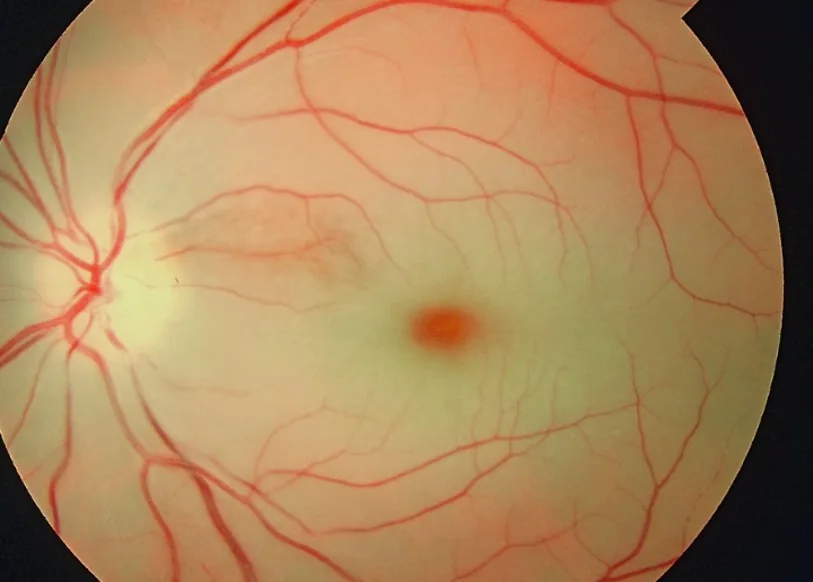
Fundus photograph, right eye.
Image by Gopal Bisht, via Wikimedia Commons. Licence: CC BY-SA 4.0.