6 Sept 2026 · Case #001
Acute ischemic stroke (middle cerebral artery)
This page works the case through in full, answer included. If you would rather reason it out yourself: Play this case
Diagnosis
Acute ischemic stroke (middle cerebral artery)
First-line treatment
IV thrombolysis and/or mechanical thrombectomy
How the diagnosis is reached
Sudden focal deficits in an older patient are a stroke until proven otherwise, and the first job is to remove the mimics that are treatable in their own right. The normal capillary glucose does most of that work, since hypoglycaemia reproduces focal deficits convincingly and would have been reversed at the bedside. Expressive aphasia with right-sided weakness places the lesion in the left hemisphere, and the rhythm strip supplies the mechanism. The QRS complexes are narrow but spaced irregularly, no two intervals alike, and not one is preceded by a P wave, only a fibrillating baseline: atrial fibrillation, which without anticoagulation throws emboli, and this one lodged in the left middle cerebral artery. Everything after that is a timing question. Onset was witnessed 45 minutes ago, so the 4.5-hour window for a plasminogen activator such as alteplase or tenecteplase is open, the CT excludes the haemorrhage that would forbid it, and a pressure of 168/92 sits under the 185/110 ceiling that has to be met before the drug runs. Thrombolysis alone is not enough here: a proximal MCA occlusion is a large clot that intravenous drug clears unreliably, and perfusion shows a small infarcted core inside a much larger territory that is still salvageable. That mismatch is what justifies retrieving the clot mechanically as well, and it is why the thrombectomy window stretches to 24 hours where imaging can still show tissue worth saving. Antiplatelets wait 24 hours after lysis.
Key clinical clues
- Sudden onset of focal deficits, clearly timed at 45 minutes
- Expressive aphasia + right hemiparesis → left hemisphere lesion
- Irregularly irregular rhythm consistent with atrial fibrillation, unanticoagulated
- Non-contrast CT excluding intracranial hemorrhage
- CT angiography showing left MCA occlusion with salvageable penumbra
The case in full
Presenting complaint
Patient history
A 67-year-old man is brought to the emergency department because of sudden difficulty speaking and weakness of his right arm. His symptoms began approximately 45 minutes ago.
Neurological examination
Physical examination
On examination he has right-sided facial drooping and reduced strength in his right arm and leg. He is awake but has difficulty producing words (expressive aphasia).
Vital signs & background
Vital signs
Blood glucose is normal. He has a history of hypertension and atrial fibrillation but is not taking anticoagulant medication.
| Parameter | Value | Reference |
|---|---|---|
| Blood pressure | 168/92 mmHg | – |
| Heart rate | 112, irregular bpm | – |
| Capillary glucose | 5.8 mmol/LRef 3.9–7.8 | 3.9–7.8 |
| Temperature | 36.8 °CRef 36.1–37.5 | 36.1–37.5 |
| SpO₂ (room air) | 97 %Ref 95–100 | 95–100 |
Bedside cardiac monitoring
ECG
He is placed on a cardiac monitor and a rhythm strip is recorded.
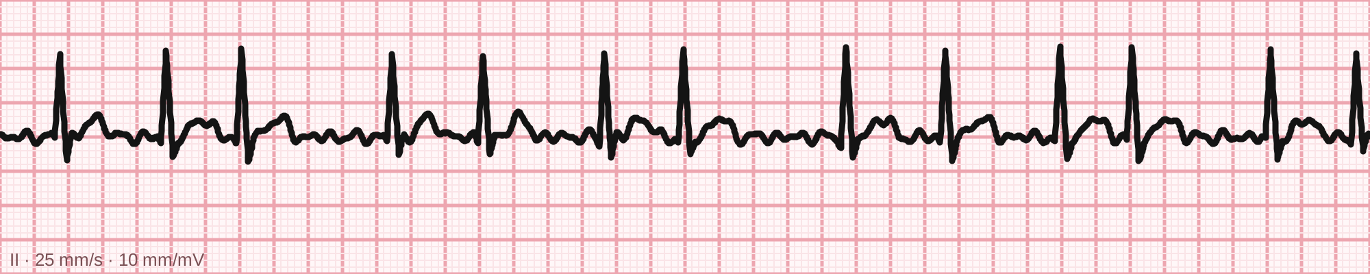
Bedside rhythm strip.
Tracing drawn for MedGuesser, not a recording from a patient.
Non-contrast CT of the head
Imaging
A non-contrast CT of the head is obtained before any treatment decision is made. There is no intracranial haemorrhage. Grey-white differentiation is already indistinct in the left basal ganglia and across the left insular ribbon, and the left middle cerebral artery is denser than its counterpart along its proximal course.
CT angiography & perfusion
Imaging
CT angiography and perfusion imaging are performed. The left middle cerebral artery stops abruptly in its proximal segment, with only sparse collateral filling beyond the cut-off. Perfusion mapping shows a small area of established infarction surrounded by a much larger territory of delayed transit that is not yet infarcted. He remains inside the treatment window.