6 Oct 2026 · Case #031
Acute angle-closure glaucoma
This page works the case through in full, answer included. If you would rather reason it out yourself: Play this case
Diagnosis
Acute angle-closure glaucoma
First-line treatment
IV acetazolamide with topical agents, then laser peripheral iridotomy
How the diagnosis is reached
Headache with nausea and vomiting frequently sends these patients down an abdominal or neurological pathway, and the diagnosis is missed until someone asks about the eye. The distinguishing features are all ocular: a red eye with circumcorneal injection, a hazy cornea from epithelial oedema, and a mid-dilated, oval, fixed pupil. That pupil is the crucial sign: a constricted pupil suggests anterior uveitis and a normal one suggests conjunctivitis, whereas mid-dilation is what occurs when the iris bunches into the drainage angle and blocks aqueous outflow. Haloes around lights are produced by corneal oedema diffracting light. The predisposing anatomy is hypermetropia, which gives a shallow anterior chamber and a crowded angle, and the two precipitants are both present here: dim light causing physiological pupil dilatation, and an antihistamine with anticholinergic activity doing the same pharmacologically. An intraocular pressure of 54 mmHg against a normal of 10 to 21 confirms it. Optic nerve damage begins within hours, so the pressure comes down now: intravenous acetazolamide to cut aqueous production, topical pilocarpine to constrict the pupil and pull the iris back out of the angle, a topical beta blocker and alpha agonist alongside, and the patient laid flat so the lens falls away from the iris. All of that is holding action. The cure is laser peripheral iridotomy, which gives aqueous a channel past the pupillary block, and the narrow occludable angle in her asymptomatic left eye is why it is done on both: the fellow eye is anatomically identical and has simply not met its trigger yet.
Key clinical clues
- Headache and vomiting where the ocular symptoms only surface on direct questioning
- Mid-dilated, oval, fixed pupil, not constricted as in uveitis
- Circumcorneal injection with a hazy cornea and haloes around lights
- Hypermetropia plus two dilating triggers: a darkened room and an anticholinergic antihistamine
- Intraocular pressure of 54 mmHg, with a narrow occludable angle in the fellow eye
The case in full
Presenting complaint
Patient history
A 66-year-old woman presents with four hours of severe right-sided headache, nausea and repeated vomiting. She was referred as a possible abdominal emergency. Pressed further, she says her right eye aches and she sees coloured rings around lights.
History and background
Patient history
Symptoms began while watching a film in a darkened cinema. She is long-sighted and wears reading glasses with strong lenses. She was started on an antihistamine for hay fever four days ago.
Ocular examination
Physical examination
Both eyes are shown below, the right eye appearing on the left of the image. The pupil does not react to light, and the globe feels hard through the closed lid compared with the left. The left eye is unremarkable.
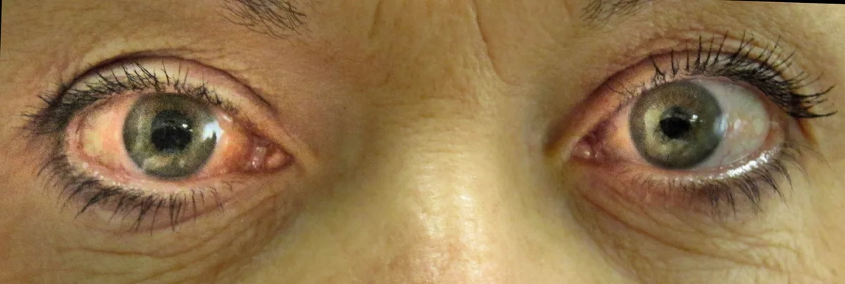
External photograph, both eyes.
Image by James Heilman, MD, via Wikimedia Commons. Licence: CC BY-SA 3.0.
Observations and bedside tests
Vital signs
Observations and visual acuity are recorded.
| Parameter | Value | Reference |
|---|---|---|
| Visual acuity, right | Counting fingersRef 6/6 | 6/6 |
| Visual acuity, left | 6/9Ref 6/6 | 6/6 |
| Heart rate | 58 bpmRef 60–100 | 60–100 |
| Blood pressure | 158/86 mmHg | – |
| Temperature | 36.7 °CRef 36.1–37.5 | 36.1–37.5 |
Abdominal assessment
Physical examination
The abdomen is soft and non-tender with normal bowel sounds. Amylase, liver enzymes, white cell count and CRP are all normal, and an erect chest radiograph shows no free air.
Ophthalmology review
Clinical course
Tonometry records an intraocular pressure of 54 mmHg on the right and 16 mmHg on the left. Slit lamp examination shows a shallow anterior chamber with the peripheral iris apposed to the cornea in both eyes. Gonioscopy of the left eye shows a narrow, occludable angle.